Your Cart is Empty


Your vocal folds — the preferred clinical term for what most people call “vocal cords” — are paired, layered mucosal tissues housed inside the larynx. According to the Cleveland Clinic, they serve three primary physiological functions: producing sound through phonation, protecting the airway during swallowing, and regulating airflow for breathing. Understanding vocal cord roles at this level of detail is what separates reactive care from genuine vocal longevity.
The three core functions, briefly:
Seek prompt evaluation if you notice: hoarseness lasting more than 2–4 weeks, sudden unexplained voice loss, difficulty breathing or swallowing, pain when speaking, or blood in your saliva. These are red flags that warrant an ENT referral, not a wait-and-see approach. The NIDCD and Harvard Health both emphasize that persistent hoarseness can signal conditions ranging from benign nodules to laryngeal cancer.
Vocal folds serve three irreplaceable roles: phonation, airway protection during swallowing, and respiration control — and preserving all three depends on consistent daily care and prompt attention to warning signs.
| Point | Details |
|---|---|
| Three primary functions | Vocal folds produce sound, protect the airway during swallowing, and regulate breathing airflow. |
| Mucosal wave is the key | A smooth, symmetric mucosal wave in the superficial lamina propria is the foundation of a clear, healthy voice. |
| Red-flag threshold | Hoarseness lasting more than 2–4 weeks requires ENT evaluation to rule out serious conditions. |
| Top prevention habits | Daily hydration, amplification use, reflux management, and scheduled vocal rest reduce chronic voice disorder risk. |
| Tmrgsolutions support | TMRG therapy kits and recovery drops are adjunctive tools for at-home maintenance alongside professional ENT/SLP care. |
The larynx sits in the midline of your neck, suspended between the hyoid bone above and the trachea below. Three cartilages define its architecture. The thyroid cartilage forms the prominent “Adam’s apple” shield at the front. The cricoid cartilage sits just below it and is the only complete cartilaginous ring in the airway — a structural fact that makes it the foundation for the entire laryngeal frame. The paired arytenoid cartilages perch on the back of the cricoid and articulate via a ball-and-socket joint, allowing them to rotate inward (adduction) or outward (abduction) to open or close the glottis. According to laryngeal anatomy research on NCBI Bookshelf, this arytenoid rotation is the primary mechanical driver of vocal fold position.
The larynx divides into three regions: the supraglottis (above the folds, including the epiglottis and false folds), the glottis (the true vocal folds and the space between them), and the subglottis (below the folds, transitioning into the trachea). The glottis is where phonation and airway protection happen.
Each vocal fold is not a simple band of muscle. It has five distinct layers, and understanding them explains why some injuries heal easily while others cause permanent voice change:
The epithelium and superficial lamina propria together form the “cover”; the deeper layers and muscle form the “body.” This cover-body model, described in PMC/NIH research on vocal fold structure, is central to understanding why mucosal health matters far more than raw muscular strength.
Male vocal folds average 17–23 mm in length; female folds average 12–17 mm. The longer, more massive male folds vibrate more slowly, producing a lower fundamental frequency. At puberty, testosterone drives rapid thyroarytenoid growth, causing the characteristic voice “break.” In older adults, the lamina propria thins and loses elasticity — a condition called presbyphonia — producing a thinner, breathier tone. Hormonal fluctuations during the menstrual cycle and pregnancy can temporarily alter fold hydration and stiffness, which is why many singers notice pitch instability at predictable points in their cycle.
Phonation begins the moment your brain signals the laryngeal muscles to bring the folds to a near-closed position — the prephonatory posture. From there, the physics take over.
The Weill Cornell Medicine Voice Center emphasizes that the “vocal cord” label is misleading precisely because it implies a guitar-string model: tighten the string, raise the pitch. Real phonation is far more nuanced. The folds are pliable tissue, not strings, and their vibration depends on the interaction between airflow and tissue elasticity — the aerodynamic-myoelastic theory.
Here is how the phonation cycle unfolds:
The mucosal wave is the clinical gold standard for assessing fold health. A smooth, symmetric wave means the cover is pliable and the layers are intact. An asymmetric, reduced, or absent wave signals scarring, stiffness, a mass lesion, or paralysis. Stroboscopy — a technique that flashes light slightly out of phase with vibration frequency to create an apparent slow-motion view — makes this wave visible in a clinical setting, as described by the Weill Cornell Voice Center. No other office test reveals mucosal wave integrity as directly.
Five intrinsic laryngeal muscles do the fine work of phonation and airway management. Each has a specific mechanical role, and each is supplied by a specific nerve branch. The table below maps the key relationships.
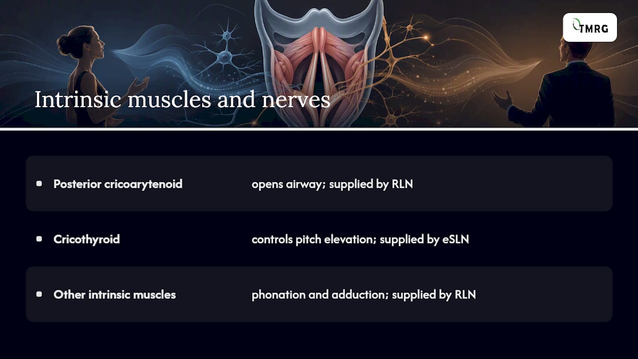
The posterior cricoarytenoid is the only muscle that opens the airway. Bilateral RLN injury — from thyroid surgery, thoracic surgery, or a tumor compressing the nerve — paralyzes both PCA muscles, leaving the folds in a near-midline position. The result is a compromised airway, not just a weak voice. Unilateral RLN injury, by contrast, typically produces a breathy, weak voice with reduced adduction on one side.
The extrinsic laryngeal muscles (suprahyoid and infrahyoid groups) do not directly control phonation but set the vertical position of the larynx in the neck. A raised larynx during swallowing protects the airway; a lowered larynx during singing opens the pharyngeal resonating space. Singers and speakers who carry chronic neck tension often have overactive extrinsic muscles that restrict laryngeal mobility and alter resonance.
The NCBI Bookshelf chapter on laryngeal anatomy confirms that the recurrent laryngeal nerve supplies all intrinsic muscles except the cricothyroid, which is why eSLN injury — less common but clinically significant — specifically impairs pitch elevation and high-note access without causing the breathy voice typical of RLN damage.
Three perceptual qualities define a voice: pitch, loudness, and timbre. Each has a distinct physiological driver, and each can be altered by specific muscle imbalances or tissue changes.
Small imbalances compound quickly. A singer who compensates for a weak CT by pressing harder with the TA may temporarily maintain pitch but at the cost of increased fold contact pressure, which raises the risk of contact lesions over time.
Pro Tip: If you notice your voice tiring faster than usual on high notes, the issue is often CT fatigue rather than general vocal overuse. Targeted breath-support work — specifically learning to increase subglottal pressure gradually rather than pressing the folds harder — can restore pitch stability without adding mechanical stress to the fold edges.
The position of the vocal folds at any moment tells a clinician a great deal about what is happening neurologically and mechanically. These are the positions you will encounter in clinical reports:
Unilateral paralysis in the paramedian position is the most common clinical scenario. The affected fold sits slightly off center; the healthy fold crosses the midline to compensate, but the closure is incomplete. Voice therapy and, when needed, injection medialization can restore adequate closure. Stroboscopy and flexible nasolaryngoscopy are the standard tools for documenting these positions and assessing whether the mucosal wave is preserved on the non-paralyzed side.
Most voice problems fall into a handful of recognizable patterns. Knowing which symptoms point to which conditions helps you decide how urgently to seek care.
Harvard Health and the NIDCD both note that many voice problems improve with conservative management once the underlying cause is identified. The key is not waiting too long to find out what that cause is.
See an ENT promptly if: hoarseness lasts more than 2–4 weeks, you have unexplained difficulty swallowing or breathing, you notice blood in your saliva, or your voice changes rapidly without an obvious cause. These presentations require laryngoscopy to rule out serious pathology. For singers and teachers, a guide to managing vocal fold ailments can help you understand the clinical pathway before your appointment.
A thorough voice evaluation typically involves more than one test, and the sequence matters. Here is what to expect:
The typical evaluation path runs from a primary care visit to an ENT referral, then to a multidisciplinary assessment with both an ENT (or laryngologist) and a speech-language pathologist. Imaging (CT or MRI of the neck and chest) is added when paralysis or a mass lesion is suspected and the cause is not obvious from the endoscopic view.
Before your appointment, bring: a voice recording from a typical heavy-use day, a written symptom timeline (when it started, what makes it better or worse), and a log of your daily voice use (hours of teaching, rehearsal, meetings). This information cuts the diagnostic workup time considerably. For professional voice users, understanding when to involve an ENT and SLP is covered in depth at Tmrgsolutions’s guide to medical professionals and vocal health.
Treatment choice depends on diagnosis, severity, and how much the voice matters professionally. Most conditions have a clear first-line approach.
Pro Tip: Absolute voice rest is rarely the right first step for most voice problems. For nodules and muscle tension dysphonia, complete silence can actually increase muscle tension and delay recovery. Guided voice therapy — using the voice in a healthy, low-effort way — is usually more effective than silence. Reserve strict rest for the immediate post-surgical period or acute hemorrhagic polyps, and always follow the SLP’s timeline.
Multidisciplinary care — ENT plus laryngologist plus SLP — produces better outcomes than either specialty working alone, particularly for professional voice users. Products from Tmrgsolutions, such as the TMRG Voice Therapy kit - Standard (TVT-S), are designed as adjuncts to this kind of structured care, supporting at-home exercise practice and daily monitoring between clinical sessions.

Vocal hygiene is not a vague concept. It is a specific, evidence-supported set of behaviors that reduce mechanical and chemical stress on the folds. Think of it the way you think about dental hygiene: consistent daily habits prevent the problems that are expensive and painful to fix later.
For warmup before a performance or heavy teaching day, a brief 5–10 minute sequence of gentle lip trills, humming on a comfortable pitch, and easy glides through your range is enough to increase fold circulation and pliability. Cooldown after heavy use — quiet humming, gentle descending glides, and a few minutes of silence — helps the folds recover from sustained vibration. For a structured daily routine, the step-by-step vocal hygiene guide at Tmrgsolutions walks through each habit in sequence.
Pro Tip: Plan your vocal-load week like an athlete plans training. Alternate one heavy-use day with a moderate or light day. On long teaching or rehearsal days, set a phone reminder for every 90 minutes as a cue to take a 5-minute voice rest. These micro-rests reduce cumulative fold contact pressure far more than a single long rest at the end of the day.
The following checklist draws on guidance from the NIDCD, Cleveland Clinic, and University of Utah Health. Each item is paired with a brief evidence note so you can verify the reasoning.
Tmrgsolutions brings over 25 years of expertise in vocal health to this guidance. For persistent or red-flag symptoms, always consult an ENT or speech-language pathologist — no checklist or product replaces a clinical evaluation.
Early vocal trouble rarely announces itself with a dramatic voice loss. More often, it shows up as a slight shrinkage in upper range, a note or two that used to come easily now requiring extra effort. Teachers notice they are projecting harder by mid-afternoon. Singers find their passaggio (register transition) feels rougher or less reliable. These are not signs of weakness or poor technique. They are the voice’s early warning system.
The most common immediate steps that help: reduce your vocal load for 24–48 hours, increase water intake, and check whether you have been sleeping poorly or fighting a mild illness (both reduce mucosal hydration and fold pliability). If the change persists beyond a week without a clear cause, schedule an ENT appointment rather than waiting for the full 2–4 week threshold. Early evaluation almost always means simpler treatment.
Many problems — early nodules, mild LPR-related inflammation, muscle tension patterns — respond very well to behavioral treatment when caught early. The window for conservative management closes as tissue changes become more established. That is the reassurance worth holding onto: most voice problems are treatable, and the earlier you act, the more options you have.
Structured clinical care from an ENT and SLP is the foundation of vocal recovery. Adjunctive products can support that foundation by making daily maintenance easier and more consistent.

Tmrgsolutions offers a range of products designed to complement — not replace — professional care. The TMRG Voice Therapy kit - Standard (TVT-S) provides guided at-home exercises and monitoring tools for singers and speakers working through a therapy program. For lighter daily maintenance or first-time users, the TMRG Voice Therapy kit - Basic offers a practical entry point. When acute hoarseness or vocal fatigue needs symptomatic support, the TMRG Loud & Clear - Classic Voice Recovery Drops (#1) are formulated for short-term soothing relief. For ongoing routine care, the TMRG Voice Clarity Trio bundles daily maintenance support into one convenient kit.
All TMRG products are adjunctive tools. If you are experiencing red-flag symptoms — hoarseness beyond 2–4 weeks, breathing difficulty, or sudden voice loss — consult an ENT or SLP before adding any product to your routine.
The following high-authority sources informed this article. Each is publicly accessible and suitable for deeper reading or clinical verification.
This article is medically referenced and written for educational purposes. For any persistent or red-flag vocal symptoms, consult a licensed ENT physician or speech-language pathologist for individual evaluation and care.
This article is general information, not a substitute for advice from a qualified doctor. Consult a qualified healthcare professional about your own circumstances before acting on anything here.
Smoking, chronic reflux, vocal overuse without adequate rest, and repeated forceful throat clearing are among the most common causes of vocal fold damage. The NIDCD notes that infections, irritant exposure, and vocal misuse all contribute to voice disorders that often improve once the underlying cause is addressed.
The main clinical positions are abducted (open for breathing), adducted (closed for phonation and swallowing), paramedian (partially closed, typical in unilateral paralysis), bowed (curved with a midline gap, common in presbyphonia), and midline-fixed (immobile at center, which can compromise the airway). Each position produces a distinct pattern of voice quality and airway function.
Singing does build coordination, breath support, and muscular endurance in the laryngeal and respiratory muscles, but the vocal folds themselves do not thicken or “toughen” the way skeletal muscles do. Improvement comes from better neuromuscular control and mucosal conditioning — which is why technique and consistent, well-paced practice matter more than sheer volume of use.
Under stroboscopy, healthy vocal folds appear pearly white or pale pink, with smooth, straight edges and a symmetric mucosal wave traveling across the cover during phonation. The glottis closes completely at the midline with no visible gap, and both folds move symmetrically. Any asymmetry, redness, lesion, or irregular wave pattern signals a problem worth investigating.
Hoarseness lasting more than 2–4 weeks without a clear cause — such as a recent cold — warrants ENT evaluation. Harvard Health notes that persistent hoarseness can indicate conditions ranging from benign nodules to laryngeal cancer, making timely laryngoscopy the responsible next step.
